If you could have one superpower, “X-ray vision” would probably be high on the list. For our Horsham veterinarians, it isn’t science fiction—it is a daily reality.
At Horsham Veterinary Hospital, we have moved beyond the darkrooms and hanging films of the past. We utilize state-of-the-art Digital Radiography (DR) to look beneath the surface of your pet’s skin.
Whether it is a limping Labrador or a cat that swallowed a hair tie, here is how this technology helps us diagnose problems in seconds.
What is Digital Radiography?
In the old days, taking an X-ray was like using a film camera. We had to take the picture, go into a darkroom, dip the film in chemicals, and wait 10 minutes to see if the image was clear.
Digital Radiography is like using a high-end digital camera. The image is captured by a sensor and appears on our computer screen almost instantly.
Why is “Digital” Better for Your Pet?
Speed: We get the image in seconds. This means less time on the table for your pet and faster answers for you.
Less Radiation: Digital sensors are incredibly sensitive, so we need much less X-ray power to get a clear picture compared to old film.
Detail: We can zoom in, adjust contrast, and measure bone angles on the computer screen. We can spot hairline fractures that might have been invisible on film.
Sharability: If your pet has a complex fracture or heart condition, we can email the digital files to a specialist surgeon or cardiologist instantly for a second opinion.
When Do We Recommend X-rays?
We use X-rays to look at “hard” things (bones) and “air” things (lungs), as well as the outline of organs.
Orthopedics: Diagnosing broken bones, hip dysplasia, or arthritis.
The “Foreign Body”: This is a classic. We look for balls, rocks, coins, and bones that have been swallowed and are stuck in the stomach or intestines.
Heart & Lungs: We can measure the size of the heart (to check for heart failure) and look for fluid or masses in the lungs (pneumonia or cancer).
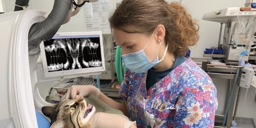
Dental: Note: We use a specialized mini X-ray machine for teeth to see the roots below the gum line.
What to Expect During Your Visit
The most common question we get is: “Does my pet need to be asleep?”
1. The Positioning
To get a diagnostic image, we need your pet to lie perfectly still in very specific positions (e.g., flat on their back or stretched out on their side). If they wiggle even a millimeter, the image will be blurry—like a bad selfie.
2. Sedation vs. General Anesthetic
Cooperative Pets: For a simple chest X-ray on a calm dog, we can sometimes do it awake with just a gentle hold.
Sedation: For painful injuries (like a broken leg) or perfect hip positioning, we usually administer a sedative. This relaxes their muscles and relieves pain, ensuring we get the shot right the first time without causing them stress.
Full Anesthetic: Usually reserved for very complex views or dental X-rays.
3. Safety
Our staff wear lead gowns and thyroid shields to protect themselves from scattered rays. Because your pet is only exposed for a split second, the radiation risk to them is negligible—the benefit of finding the problem far outweighs the risk.